
Endodontic surgery About 9-minute read
Apicoectomy Procedure Steps and Recovery Timeline
An apicoectomy, also called root-end surgery, treats persistent inflammation or infection around a tooth root after root canal treatment when retreatment is unsuitable or unlikely to resolve the cause. Knowing the sequence—from examination and consent to sutures and follow-up—can make the experience feel more predictable.
Clinically reviewed information: This educational guide describes common practice. Your surgeon’s instructions take priority because anatomy, medical history and surgical findings vary.
Apicoectomy at a glance
Purpose
Remove infected or inflamed tissue at root tip, trim a small portion of root end, and seal canal end where indicated.
Anaesthesia
Usually local anaesthesia; individual plans differ.
Healing
Soft-tissue comfort often improves over days; bone healing takes longer and is checked over months.
Exact appointment length and recovery depend on tooth position, access, surgical complexity and whether other care is needed. These are individual estimates made after examination, not fixed promises.
When clinician may consider root-end surgery
Inside each tooth, root canal treatment cleans and seals canal system. Occasionally inflammation persists or returns near root tip despite appropriate treatment. Reasons can include complex canal anatomy, a persistent lesion, a blockage that prevents safe retreatment, or an existing restoration that makes removing and replacing prior work risky. An apicoectomy approaches problem from root end rather than through crown.
It is not automatic next step after every failed root canal. Clinician evaluates symptoms, tooth restorability, periodontal support, quality of existing root filling, bite, and imaging. Options may include monitoring, non-surgical root canal retreatment, apicoectomy, or extraction when tooth cannot predictably be retained. A second opinion from an endodontist or oral surgeon may help for complex cases.
The goal is to remove diseased tissue and seal route of leakage while preserving a restorable tooth where reasonable. No procedure guarantees healing. A clinical and radiographic follow-up plan helps assess outcome.
Apicoectomy preparation and examination
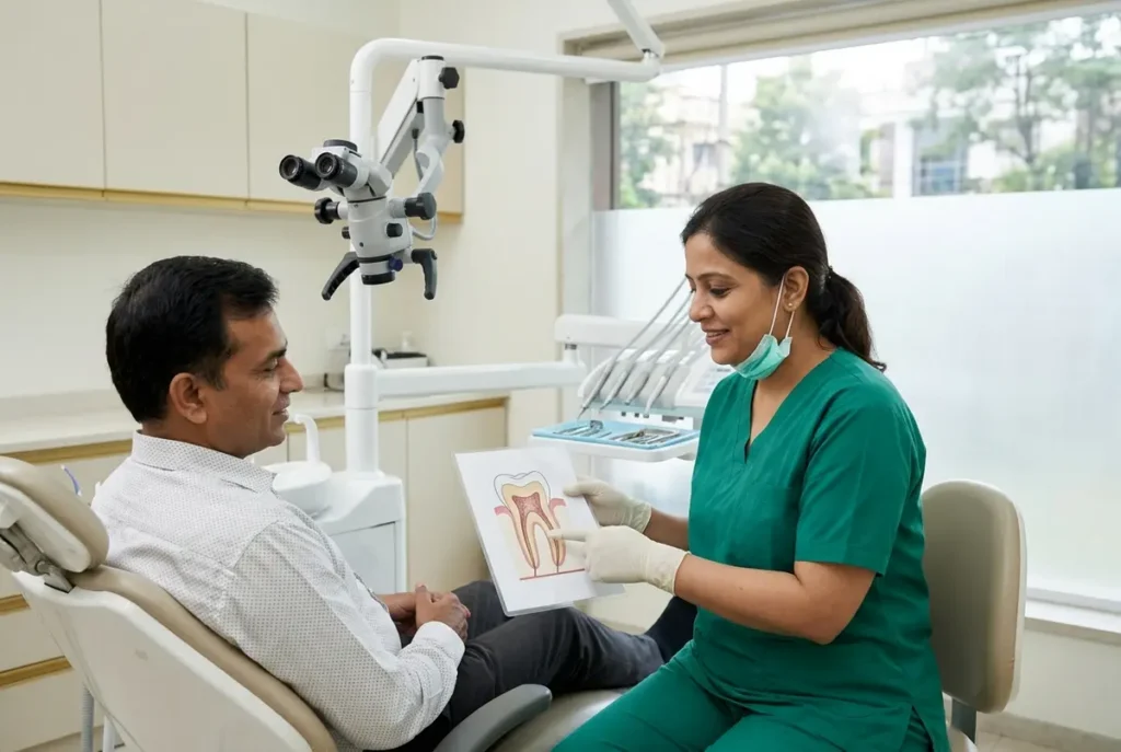
Appointment begins with history and focused assessment. Tell dental team about prescribed and non-prescribed medicines, allergies, medical conditions, previous reactions to anaesthesia, pregnancy, smoking, and any bleeding or immune concerns. Do not stop prescribed medication unless prescriber or treating clinician advises this. If sedation is planned, follow instructions about food, drink, escort and driving; local-anaesthetic-only cases may have different guidance.
Clinician checks tooth, gums, bite and nearby structures. Periapical dental X-rays are common; cone-beam CT may be recommended when three-dimensional detail could change planning, especially near nerves or sinus. Imaging is selected for clinical need, not used automatically. The team reviews tooth position, root anatomy, lesion extent and restoration before discussing plan.
Before consent, ask what diagnosis supports surgery, alternatives, likely benefits, limitations, material risks, expected appointments and follow-up. Risks can include post-operative pain, swelling, bruising, bleeding, infection, delayed healing, persistent symptoms, damage to nearby structures, or need for further treatment. Risks vary by site. Saluja Dento Max Fac Centre in Kanpur describes its care as examination- and imaging-led, with treatment options and consent discussed before procedures.
Root-end surgery steps: what happens in the procedure
Sequence varies by tooth and surgical approach. Microsurgical instruments and magnification may be used to improve visibility and precision. Here is a general outline, not a substitute for the operating clinician’s explanation.
01 · Anaesthesia and access
Local anaesthetic numbs area. Once numbness is confirmed, surgeon makes a small incision in gum and gently reflects tissue to reach bone over root tip. Patient may feel pressure or vibration, not sharp pain; report pain immediately.
02 · Root-tip access
A small opening through bone exposes root end and surrounding inflamed tissue. Surgeon removes affected tissue for treatment and, when appropriate, laboratory examination. Care is taken to protect adjacent roots, nerves and sinus.
03 · Resection and seal
Surgeon trims a small portion of root tip, inspects cut surface and prepares a small root-end cavity. A biocompatible filling material seals canal end. This seal aims to reduce pathways for bacteria; exact technique and material depend on case.
04 · Closure and instructions
Area is irrigated, tissue repositioned and gum closed with sutures. Surgeon may take a post-operative image, then explain cold-pack use, oral hygiene, food choices, medication and review timing before discharge.
During surgery, draping and instruments can make conversation difficult; agree on a hand signal to pause. Pressure, pulling and vibration are common sensations. Tell clinician if you feel sharp pain, become faint, or need a break.
Apicoectomy compared with non-surgical retreatment
These approaches solve different access problems. Clinician recommends option based on cause of persistence, tooth structure and feasibility—not a universal ranking.
| Factor | Root canal retreatment | Apicoectomy |
|---|---|---|
| Access route | Through crown and existing restoration into canals. | Through gum and bone to root tip. |
| May suit | When canal can be accessed and cleaned or sealed again. | When problem remains at root end and conventional retreatment is unsuitable or insufficient. |
| Recovery focus | Tooth and restoration; surgical wound is not typical. | Gum incision, swelling, tenderness and sutures. |
| Main planning question | Can source be corrected from inside tooth? | Can root end be safely reached and sealed while preserving tooth? |
Healing after apicoectomy: recovery milestones
Recovery is a range, not a countdown. Mild pain, swelling, bruising or tenderness can occur after oral surgery. Swelling may be most noticeable during first few days and then settle, but response differs. Follow written instructions from your treating team rather than using timeline below as a diagnosis.
Day of procedure
Rest; use cold pack externally in short intervals if instructed. Avoid strenuous activity. Keep gauze in place only as directed. Wait until sensation returns before chewing, and avoid hot food or drink while numb.
First 24–72 hours
Choose soft, cool or lukewarm foods; chew away from surgical site. Do not poke wound, vigorously rinse, spit forcefully or smoke. Use prescribed medicines exactly as directed. Pain should be manageable and trend gradually better.
Following days
Resume gentle brushing, carefully avoiding incision at first. Rinse only as advised; do not pull at sutures. Stitches may be absorbable or require removal, often at a review around a week, but timing depends on clinician and wound.
Soft tissue usually settles sooner than underlying bone. Bone repair cannot be judged by pain alone: radiographic healing develops over months, and clinician may schedule later imaging based on initial lesion and findings. Attend both early wound review and longer-term check when advised.
Sutures, follow-up and practical aftercare
Keep the surgical area clean without brushing directly over fresh stitches. Follow surgeon’s directions for mouth rinses and brushing; advice can differ with incision location. Do not use a straw or disturb wound if told to avoid suction. Drink water and eat soft foods that do not require hard chewing. Avoid smoking and alcohol during early healing, as these can interfere with recovery and medicines.
Take any prescribed pain relief or other medication according to label and clinician’s instructions. Do not add aspirin or combine medicines without checking, especially if you have medical conditions or take blood thinners. Antibiotics are not automatically needed after every procedure; use them only when prescribed and complete course as directed. Ask clinic if dose is missed or side effect occurs.
At follow-up, clinician checks wound closure, swelling, tenderness and sutures; non-dissolving stitches may be removed. You may be asked about numbness, drainage or bite discomfort. Longer-term reviews assess symptoms and bone healing on imaging. Keep the tooth’s restoration maintained: a sound coronal seal and routine dental care remain important after root-end treatment.
Warning signs: when to contact dentist promptly
Contact treating clinician promptly if pain or swelling becomes markedly worse instead of improving, bleeding continues despite pressure as instructed, fever develops, pus or persistent bad taste appears, wound opens, or medication causes a concerning reaction. New or worsening numbness, difficulty opening mouth, or a change in bite also warrants advice. Breathing or swallowing difficulty, rapidly spreading facial or neck swelling, or severe allergic symptoms require emergency medical care.
If unsure, call clinic and describe date of surgery, symptom onset, temperature if measured, medicines taken and whether symptoms are worsening. Do not wait for scheduled review when symptoms are escalating.
Apicoectomy procedure and recovery FAQs
Will apicoectomy hurt?
Local anaesthesia is used in many cases, so sharp pain should not be expected during treatment. Pressure, movement and vibration can be felt. After numbness fades, tenderness and swelling are possible; clinician advises suitable pain relief based on your health and medicines.
How long until stitches come out?
Some sutures dissolve; others need removal at a follow-up, commonly planned within about a week depending on surgical site and material. Do not pull or cut them yourself. Confirm your specific review date with surgeon.
When is bone fully healed?
Soft-tissue comfort may improve over days, but bone healing takes substantially longer and is assessed clinically and, when appropriate, with follow-up radiographs over months. Timing varies by lesion size, site and individual healing.
Can I return to normal work next day?
Some people resume routine activity soon; others need more rest, particularly after complex surgery or sedation. Avoid strenuous exercise initially and follow surgeon’s advice. Sedation requires separate restrictions on driving and supervision.
Discuss apicoectomy options in Kanpur
Persistent symptoms after root canal treatment need an examination, not guesswork. Saluja Dento Max Fac Centre provides dental and oral-surgical care in Kanpur, with consultation, imaging when indicated, treatment discussion and follow-up planning.
Clinic: 111A/20A, G.T. Road, near Coca Cola Chauraha, Ashok Nagar, Kanpur – 208012, Uttar Pradesh.
Hours: Monday–Saturday, 10:00 AM–7:30 PM; Sunday, 10:00 AM–2:00 PM.
Related dental reading
Explore root canal care and oral and maxillofacial surgery at Saluja Dento Max Fac Centre.
Apicoectomy Procedure Steps and Recovery Timeline
Learn apicoectomy procedure steps and recovery timeline, from imaging and root-end surgery to sutures, healing milestones, follow-up care…

Pain or Swelling After Root Canal: Causes & Next Steps
Pain or swelling near a tooth after root canal treatment? Learn common causes, warning signs, what follow-up checks…

What Is an Apicoectomy? Root-End Surgery Explained
Learn what an apicoectomy means, why infection may persist after root canal treatment, how root-end surgery works, and…