
Kanpur, Khoon Se Bhare Daant “Chhoti Baat” Nahi Hain
The stage of gum disease where teeth do not wobble yet — but they have already started planning their exit. Here is the plain-language, no-nonsense guide to pyorrhoea treatment in Kanpur, written for the families who keep saying “chalega”.
Oral Hygiene & Preventive Care • 14 min read • Updated 2025
There is a specific moment in periodontal disease that almost no family recognises: the tooth is still firmly in the jaw, still chews, still looks “normal” in a mirror — and yet the bone that holds it has already started dissolving. This is the stage most people in Kanpur treat with a stronger toothpaste, a herbal khoon-badhi dawa, or simply by changing the brushing side. By the time a tooth actually falls out, the damage behind it is often 8–12 years old. Understanding this quiet middle stage is exactly what good pyorrhoea treatment in Kanpur is built around: catch the disease while the tooth is still savable.
Brush Karte Hi Khoon? The 45-Year-Old Uncle Who Lost Six Teeth
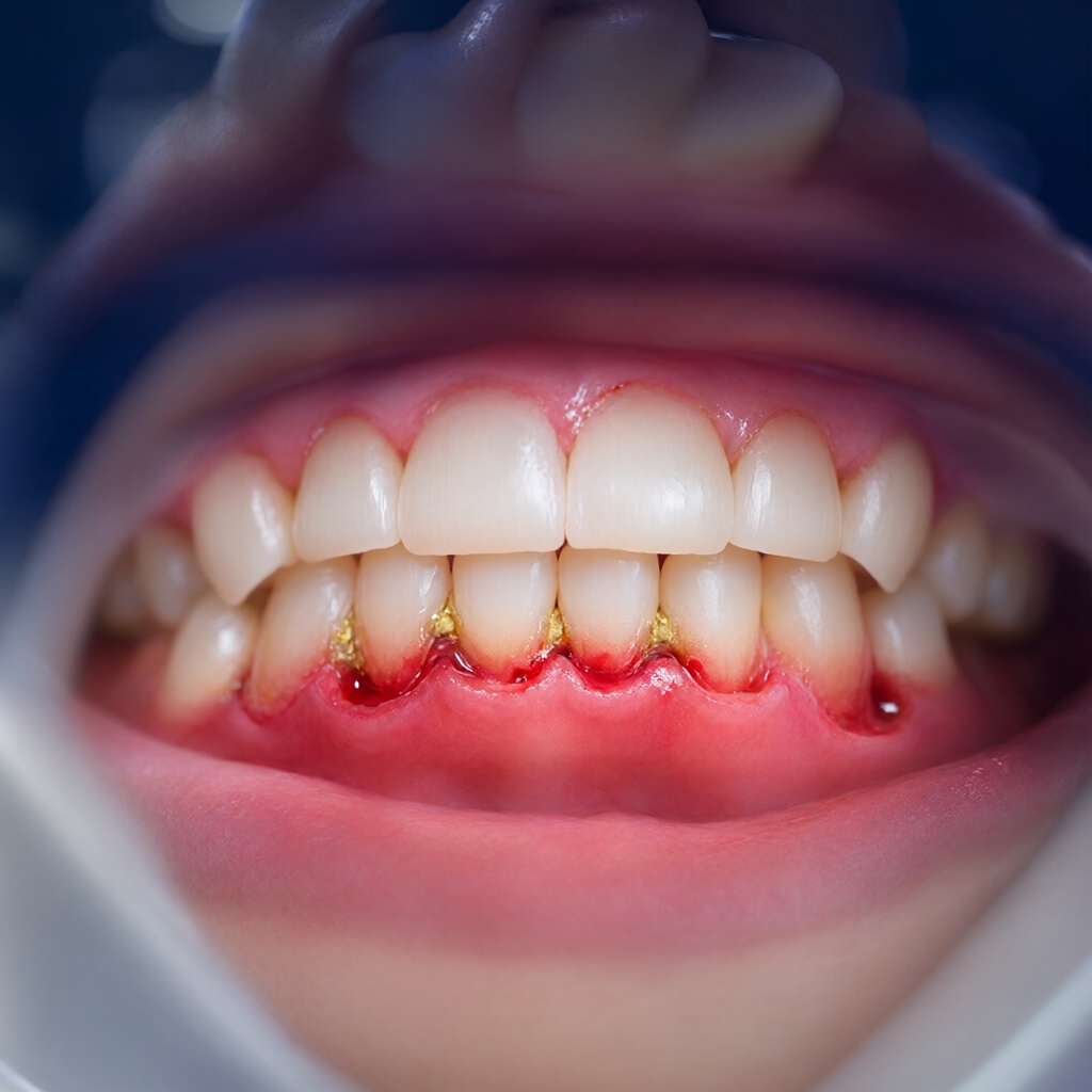
He was 45, a shopkeeper in Kanpur Cantt, and he came in complaining that his toothbrush was “cutting” him. Every morning, pink foam. He had switched to a herbal gum-powder on WhatsApp advice, then to a “khoon nikalne” tablet from the medical store, then to neem datun he chewed after every meal. Four months of effort. The bleeding had reduced — because the gums had stopped being inflamed at the gum edge, not because the disease had retreated. Under the calculus, the pocket measured 7 mm with bone loss visible on the radiograph. Six teeth were already at Grade II mobility.
This is the standard pattern in periodontal clinics, and it is worth naming clearly: visible bleeding is not the disease, it is the alarm. Plaque-induced gingivitis — the reversible stage — bleeds because the gum margin is inflamed and shallow. Once bacteria migrate under the gum line and form a true periodontal pocket, the tissue bleeds deeper, the bone begins to resorb, and the pocket wall becomes thin enough that you can literally see through it on an X-ray.
The central promise of this article: if the tooth has not started to move, you are still inside the window where treatment works predictably. Once mobility appears, you are no longer treating a gum problem — you are negotiating with bone loss.
Those numbers are not scare statistics; they are the reason periodontitis is now formally graded in stages (I–IV) and grades (A–C) by the 2017 World Workshop classification, and why the European Federation of Periodontology (EFP) S3 guideline treats it as a step-wise protocol rather than a single “cleaning” appointment.
The Real Map of the Mouth: Gum, Pocket, Tartar & the Silent 10-Year Disease
To understand pyorrhoea treatment, you need four words: gum, pocket, calculus, bone. They are a sequence, not a list.
- 1. The gum (gingiva): A living seal. It is designed to wrap the tooth like a tight collar. A healthy collar hugs the tooth with a sulcus depth of only 0.5–3 mm and does not bleed when you brush it.
- 2. The pocket: When the gum seal fails, the sulcus deepens into a measurable periodontal pocket. Once it passes 3–4 mm, a toothbrush, floss, and even a mouthwash physically cannot reach the bottom. It becomes a protected, anaerobic habitat — the perfect incubator for gram-negative bacteria and their endotoxins.
- 3. Calculus (tartar): Mineralised plaque. It forms within roughly 4–12 weeks of inadequate cleaning and becomes porous, cementum-anchored. Calculus is a physical barrier: even a skilled brush cannot touch the root surface underneath it.
- 4. The bone: Bacterial toxins and the body’s own exaggerated immune response trigger osteoclast activity. Attachment is lost, the bone height drops, and the root begins to look “long.” This is the diagnosis: clinical attachment loss — the single most important measurement in periodontology.
Chart 1 — How deep the pocket gets as pyorrhoea advances
Healthy sulcus 0.5–3 mm. Beyond 4 mm, no home device reaches the bottom. Read this as a “point of no return” ruler.
Chart 2 — The silent 10-year disease
Modelled loss of attachment (mm) over a decade in an untreated patient. Notice the flat early years — that is why nobody notices — followed by an accelerating final phase.
5 Warning Signs Kanpuriya Ignore — and the One That Means Run Today
Stage I periodontitis is, by definition, asymptomatic. You feel nothing. That is the whole problem. The five signs below are what patients actually notice, usually years too late. Treat the first four as “book a screening,” and treat the fifth as “do not brush this tooth until you have been examined.”
| Warning sign | What is actually happening | Urgency |
|---|---|---|
| 1. Persistent bad breath | Volatile sulphur compounds from anaerobic pocket bacteria. Mouthwash masks it for 20 minutes; it is back within the hour. | Screening |
| 2. Blood in the foam after brushing | Gingivitis or an inflamed pocket wall. Bleeding on probing is an active inflammation marker, not a mechanical injury. | Book this month |
| 3. A gap or khaali jagah after eating | Food packing into a pocket where gum tissue has receded. The gum no longer fills the embrasure. | Book this month |
| 4. Teeth that look “daaler” (longer, thinner) | 1–2 mm of recession means roughly 1–2 mm of bone already gone. Black triangles appear between teeth. | Within 4 weeks |
| 5. One tooth that moves when you push it | Mobility Grade I–III. The periodontal ligament and supporting bone can no longer resist normal chewing forces. | Today |
| 6. Dahi, dahalna, burning on eating | Dentin exposed by recession; root sensitivity. Sometimes a sign of an abscess hiding under the gum. | Urgent |
| 7. Metallic taste / constant urge to rinse | Bacterial biofilm load plus possible dry mouth, which dramatically accelerates the whole cascade. | Screening |
Chart 3 — What Kanpur periodontal patients report, ranked
Patterns observed across adult screening visits: bleeding on brushing dominates, mobility is chronically under-reported until it is late.
The clinical reality is that mobility is a late sign. By the time a patient notices a tooth moving, the EFP staging will almost always place them in Stage III or IV. That is precisely why the title of this piece says teeth do not need to move before the disease is serious — the pocket depth, not the wiggle, is the real scoreboard.
Sugar, BP, Medicines & Dry Mouth: The Real Drivers Behind Kanpur’s Pyorrhoea Cases
Ask any periodontist what drives progression in Indian patients and the answer is rarely “we do not brush.” It is a combination of modified risk factors, several of which are also very Kanpur: paan with kharra, gutka and khaini, bidi smoking, uncontrolled diabetes, and polypharmacy for blood pressure.
Chart 4 — Relative contribution of major risk factors
Multiplicative effect of local and systemic risk factors on periodontal progression. Tobacco and diabetes remain the two highest-yield targets for intervention.
Sugar and refined carbohydrates. Every gram of fermentable sugar that lingers for 20+ minutes drops the plaque pH below the critical 5.5 threshold and pushes the ecosystem toward acidogenic, aciduric organisms. In a city with an enormous sweet-shop and roadside kulfi culture, this is a daily exposure, not a weekly one.
Tobacco, gutka, khaini and bidi. Smokers are consistently shown to progress faster and respond less well to non-surgical therapy; smokeless tobacco causes vasoconstriction that masks bleeding (so smokers “don’t bleed” and yet have deeper pockets), stain, and impair fibroblast healing. Kharra and paan are not a substitute for cleaning.
Diabetes. The relationship is bidirectional. Poorly controlled diabetes (high HbA1c) triples the risk of attachment loss, and periodontal inflammation makes glucose control harder. This is why a Stage III patient with diabetes is treated as a systemic case, not just a cleaning appointment.
Medications that swell the gums. Calcium-channel blockers (most commonly amlodipine), phenytoin, and cyclosporine can produce drug-induced gingival overgrowth. Suddenly the pocket is not a bacteria problem — it is a plaque-retentive shape problem that cannot be solved by brushing alone and often needs flap reduction plus a physician review of the drug.
Dry mouth (xerostomia). Saliva is the mouth’s natural buffering and antibacterial system. Dehydration, AC-room hours, bidi, antihistamines, antidepressants, and anxiety all reduce salivary flow. Without saliva, bacterial counts rise dramatically, calculus forms faster, and mucosa becomes ulcer-prone. For many patients, addressing dry mouth is a genuine part of pyorrhoea treatment in Kanpur, not an afterthought.
| Risk factor | Mechanism | What your dentist will do about it |
|---|---|---|
| Tobacco / gutka / khaini | Vasoconstriction hides bleeding; impairs collagen repair | Counselling + staged therapy + longer recall interval |
| Uncontrolled diabetes | AGE-mediated collagen cross-linking, immune dysfunction | Coordinate with physician; periodontal therapy as part of glucose care |
| High BMI / sedentary lifestyle | Adipokines drive pro-inflammatory state | Lifestyle counselling alongside debridement |
| Amlodipine & similar drugs | Drug-induced gingival enlargement | Physician review for alternatives; flap surgery if severe |
| Dry mouth | Loss of buffering, buffering pH drop, faster calculus | Xylitol gum, sialogogues, hydration, alcohol-free rinses |
| Irregular dental visits | Calculus never gets removed; pockets deepen | 3-monthly supportive periodontal care after therapy |
The Stage-wise Game Plan: Scaling, Deep Cleaning, Flap Surgery & the Toothpaste Myth
Modern periodontal therapy is not a single procedure. It is the EFP S3 “four-step” protocol, and every stage you are in tells us which step you start at. This is what a genuinely staged pyorrhoea treatment in Kanpur plan looks like.
Step 1 — Behaviour change and supragingival biofilm control
For Stage I and early Stage II, this step alone can be the whole treatment. It means a professional supra- and subgingival cleaning, a demonstrated brushing technique, interdental cleaning, and tobacco cessation advice. Re-evaluation in 6–8 weeks decides whether you can stop here. Pocket depth and bleeding-on-probing are the two numbers that decide it.
Step 2 — Subgingival instrumentation (scaling and root planing / deep cleaning)
This is the gold-standard non-surgical therapy. Under local anaesthetic, an ultrasonic scaler plus hand instruments are used to remove the bacterial biofilm and calculus from every root surface to the bottom of the pocket, and the root is smoothed so new tissue can attach. Evidence consistently shows meaningful pocket reduction (typically 1–2 mm) and a high proportion of pockets returning to a maintainable depth. It is uncomfortable; it is not agony. Most patients report the first 24 hours as tender, with mild gum shrinkage over the next weeks that makes the teeth look better, not worse.
Step 3 — Periodontal surgery for residual deep pockets
If, after 3 months, pockets of 4 mm+ still bleed, or there are sites 6 mm+ that did not respond, the EFP guideline points to access flap surgery (and, where regeneration is possible, regenerative procedures using bone-graft material and membranes). The goal is not cosmetic flap — it is to create a contour the patient can actually clean, and to reduce the pocket to a depth hygiene can maintain. A flap procedure for a single deep, bleeding 6 mm site is a small surgery with a large payoff.
Step 4 — Supportive periodontal care forever
This is the step most clinics skip and patients resent paying for — and it is the step that decides whether the teeth survive. Recall every 3 months, pocket charting, re-cleaning, and home-technique reinforcement. Periodontal patients are not “cured”; they are managed. Indefinitely.
Chart 5 — Expected pocket-depth reduction by treatment step
Illustrative mean reduction in pocket depth from baseline, by therapy phase. Scaling does the heavy lifting; surgery cleans up the stubborn sites; maintenance protects the gain.
The “toothpaste se chalega” myth, dismantled
Myth 1: “A gum-strengthening paste will fix it.”
Truth: no toothpaste reaches 5 mm. Toothpaste works on the gum margin; pyorrhoea lives in the pocket floor. A good paste with fluoride + an anti-plaque agent is still essential — just not curative beyond Stage I.
Myth 2: “Charcoal / herbal powder bleaches and cleans.”
Truth: charcoal powders are abrasive with no clinical advantage, and many herbal khoon-badhi powders contain contaminants. Neither removes subgingival calculus.
Myth 3: “Scaling makes the gap between teeth bigger.”
Truth: scaling removes swollen tissue. The apparent gap is the true gum level returning. The tooth was never longer — the gum was just covering more of it.
Myth 4: “It will go away on its own.”
Truth: gingivitis is reversible; established periodontitis is not. Only the progression can be arrested, and only with mechanical removal of calculus plus maintenance.
Gum Care at Home: Brushing, Interdental Brushes, Neem Datun & 7 Rules
Home care cannot remove an existing 7 mm pocket, but it absolutely decides whether the surgery you paid for survives five years or two. Cochrane reviews consistently show that interdental brushes reduce gingivitis more effectively than floss wherever they fit, and electric toothbrushes remove plaque better than manual brushing. The technique, not the brand, is the differentiator.
Table — The 7 non-negotiable rules for gum health in Kanpur
Print this. It is the difference between a stable mouth for 20 years and a denture by 55.
Rule 1 — Technique over force. 45–60 seconds, twice daily, with a soft bristle head held at 45° to the gum line in small vibrating movements. Scrubbing side-to-side like a saw causes recession and bleeding without removing more plaque.
Rule 2 — Interdental brush every night. Choose the size that slides through the contact point with slight resistance, then move in and out 3–4 times per space. Behind the last molar, use a single-tuft brush. For tight contacts, a 0.6–0.8 mm brush will outperform floss dramatically.
Rule 3 — Neem datun: partly true. The datun does scrub plaque mechanically and has some demonstrated antibacterial activity, which is a genuine plus. The problems are practical: most families use it irregularly, the stem is often too thick (causing gum cuts and recession), and the habit is usually an addition to brushing, not a replacement. Use a slim, clean datun occasionally; keep the brush as the primary tool.
Rule 4 — Fluoride toothpaste + interdental care. Use a fluoride toothpaste (1350–1500 ppm) twice daily. If you have recession, an active cavity, or dry mouth, a higher-fluoride prescription paste helps remineralise exposed root surfaces.
Rule 5 — Manage dry mouth deliberately. Sip water through the day, chew sugar-free xylitol gum, avoid tobacco and alcohol-based rinses, and tell your dentist about every medicine you take.
Rule 6 — Short-course chlorhexidine only when prescribed. 0.12–0.2% chlorhexidine for 7–14 days is useful after surgery or in acute infection, but long-term use stains teeth, alters taste, and disrupts the oral microbiome.
Rule 7 — Never skip the 3-month recall. A maintained periodontal patient with an immaculate home routine can still have calculus. Three-monthly cleaning is cheaper than one implant, by an order of magnitude.
“Nobody loses a tooth to pyorrhoea overnight. They lose it to a pocket that was measured once, dismissed once, and then not measured again for nine years.”
What Kanpur Patients Should Do This Month
1. Do not wait for the tooth to move. Mobility means you are already in Stage III. Pocket depth and bleeding scores are the early-warning system, and they are measured in five minutes with a periodontal probe.
2. Get a proper gum screening, not a “cleaning.” Ask for pocket charting, bleeding index, and a radiograph at the affected sites. Without those three numbers, no treatment plan is truly personalised.
3. Follow the steps. Cleaning → deep cleaning → surgery only if needed → 3-monthly maintenance. The stage you are in determines where you enter, and entering at the wrong step is how people end up with a full-arch implant plan they did not need at 40.
4. Fix the drivers. Gutka, kharra, bidi, unchecked sugar, untreated diabetes, and dry mouth are all modifiable. In practice, pyorrhoea treatment in Kanpur succeeds long-term only when the patient changes at least one of these — otherwise the same tooth returns every eighteen months.
5. Understand the prognosis honestly. Gingivitis is reversible. Stage I–II periodontitis can often be stabilised for years. Stage III–IV with Grade C progression (rapid loss, often smoker or diabetic) has a genuinely guarded long-term outlook, and early intervention is what changes that forecast. Stage-wise, affordable, protocol-driven gum bleeding treatment in Kanpur is a decision about timing: the earlier you start, the more of your own teeth you keep.
Gum Screening, Pocket Charting & a Written Plan — in One Visit
If any of the five warning signs above apply to you or someone at home, book a periodontal screening. You leave with your pocket numbers, an X-ray-based stage diagnosis, and a stage-wise treatment plan with a written cost estimate — not a vague “cleaning khwa lo” advice. Message us on WhatsApp for the earliest slot, or walk in during clinic hours.
Book a Gum Screening See All Dental ServicesRelated Articles
Clear Aligners vs Braces for Adults in Kanpur: A Full Guide
Cosmetic & Aesthetic Dentistry • 11 min read Clear Aligners vs Braces for Adults in Kanpur: How Each…
What Is Osseointegration in a Dental Implant? | Kanpur Guide
Dental Implants · Implantology · 9 min read What Is Osseointegration — and Why It Decides Whether Your…
What Is Osseointegration? How a Dental Implant Fuses With Bone
Dental Implants • 12 min read What Is Osseointegration? Why a dental implant must literally grow into the…