Most people in Kanpur meet their first wisdom tooth problem somewhere between 17 and 24 — a dull ache at the back of the jaw, a bad taste that will not go away, or a sudden swell under the angle of the mandible. Some of those teeth can stay exactly where they are for a lifetime. Others quietly damage the neighbouring second molar for years before anyone realises it. This guide walks through wisdom tooth extraction in Kanpur end to end: how to know removal is genuinely indicated, what imaging reveals, how the procedure is planned and performed, what recovery really feels like in humid Kanpur weather, and — just as importantly — when a surgeon should tell you to wait.
1. Why Third Molar Problems Are Rising Across Kanpur
Third molars are the last teeth to erupt, usually between 16 and 21, and they are the only teeth that are routinely absent. Around half of all people develop at least one wisdom tooth; many have all four. The universal problem is space. Evolutionary jaws were smaller than modern diets produce, so a third molar usually arrives into a jaw that is already full — and then tilts, rotates or stops halfway.
Three local factors accelerate the problem in Kanpur. First, pericoronitis: when a tooth breaks through the gum only partially, the flap of gum over it forms a trap. Food lodges there, brushing cannot reach it, and within days you have a localised, exquisitely painful infection. Second, the second molar as a victim: a mesioangular lower third molar presses on the distal surface of the second molar, stripping bone and gum attachment and opening a pocket that is impossible to clean. Caries often starts in the adjacent second molar rather than in the wisdom tooth itself — the tooth you came to complain about is healthy, and the one you are ignoring is decaying. Third, follicle pathology: a dentigerous cyst or an enlarged dental follicle around the crown can quietly resorb bone for years without producing a single symptom.
The most important clinical message from oral surgery literature is the AAOMS line that patients repeat back to their families: “pain-free does not mean disease-free.” A completely buried, symptomless wisdom tooth can still be destroying the bone that supports the second molar. That is precisely why “I will get it removed only if it hurts” is a dangerous default — and why patients in Kanpur often arrive at an oral surgery centre years later with a more complicated, and more delicate, surgical plan than they needed in their twenties.
Delay is usually driven by three anxieties: fear of the chair, fear of the swelling, and fear of the paperwork. All three are legitimate concerns that a good consultation should address honestly. But the cost of waiting is measurable — root resorption, a second molar that suddenly needs root canal treatment, an infection that has spread into the fascial spaces of the neck, or a follicular cyst that has turned a simple 30-minute procedure into a staged one. Understanding the trade-off is the first step of making a calm, informed decision.
Where patients present most often (clinic case mix)
Common reasons third molars are removed
Relative frequency of indications seen in a maxillofacial practice — the recurring infection group dominates because it is the group that hurts.
Indicative distribution of clinical presentations, not a statistical survey. Your surgeon will classify your case individually.
2. How to Tell If Your Wisdom Tooth Actually Needs Removal
Extraction is not automatic for every third molar. It becomes the right call when there is a specific, demonstrable reason. The diagnosis almost always combines three inputs: what you describe, what the surgeon sees and probes, and what the imaging shows.
Symptoms that point towards surgery
- Pain at the back of the jaw that keeps returning. Episodic pain around the third molar, especially when chewing or opening wide, is the classic presentation of pericoronitis.
- Swelling in the gum flap or on the cheek. A tender, warm, red pocket behind the second molar that bleeds easily when you poke it.
- Bad taste or persistent halitosis. Infection in the follicle produces a metallic or foul taste that brushing does not fix.
- Jaw stiffness and reduced opening. If a normal yawn or a bite of food now hurts, the infection has reached the surrounding muscles.
- Pain radiating to the ear, temple or opposite side. Referred pain from the lower third molar is frequently mistaken for an ear infection or a sinus issue.
- Decay appearing on the second molar. New caries on the distal surface of a healthy back tooth is one of the strongest arguments for removing the wisdom tooth next to it.
What imaging actually changes
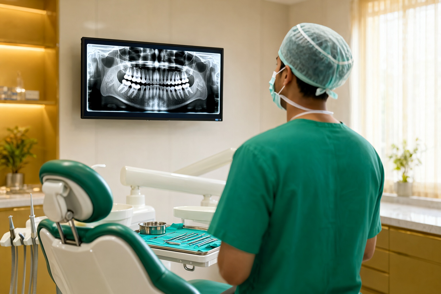
A single periapical film is not enough for a lower third molar. A digital panoramic radiograph (OPG) gives the whole arch in one low-dose view and shows angulation, impaction depth, relation to the inferior alveolar canal, the follicular space and the condition of the second molar. Where the relationship to the nerve is close, a CBCT scan gives a true 3D map — and it is the single most useful test for deciding whether a deeply buried tooth can be removed safely, or whether the safer option is to leave it in place and monitor. Ask to see your film and have the surgeon point out what is being read from it; a patient who can see the canal–root proximity understands the plan far better than one who only hears a duration.
Extraction or watchful waiting? A decision grid
| Finding | What it usually means | Usual plan |
|---|---|---|
| Fully erupted, cleansable, healthy, no pathology | Normal functional third molar | Monitor at routine visits |
| Partially erupted with a gum flap | Pericoronitis risk, cleaning difficult | Extraction, often in the same visit as the consultation |
| Horizontal / mesioangular impaction contacting the second molar | Distal bone loss and caries risk on the neighbour | Extraction before damage advances |
| Vertical impaction, deep, roots close to the inferior alveolar canal | Elevated nerve-injury risk | CBCT assessment; monitor if asymptomatic, remove if pathological |
| Enlarged follicular space or radiolucency | Cyst or follicular pathology | Prompt surgical removal with specimen for histopathology |
| Recurrent infection despite antibiotics, or spreading swelling | Antibiotics manage it, they do not remove the cause | Definitive extraction once acute infection is controlled |
3. What the Procedure Involves at a NABH-Accredited Oral Surgery Centre
Third molar removal ranges from a straightforward, forceps-assisted removal of a fully erupted tooth to a surgical procedure where gum is lifted, bone is drilled away, the tooth is sectioned into pieces and lifted out, and the gum is re-stitched. Both happen under local anaesthesia, and in both cases the patient usually hears and feels less than they expect — pressure, vibration and tugging, not drilling pain. For anxious patients, a premedication or a sedated technique changes the experience entirely.
The clinical setting matters more than patients realise. At an accredited centre, the procedure is preceded by a documented medical history review, a documented consent discussion that includes nerve risk, and a planned backup for an unexpected finding. Sterilisation follows NABH-mandated protocols, instruments are traceable by batch, and there is a documented emergency plan including the ability to manage a bleeding episode or a difficulty with the airway. These are exactly the standards a patient should expect from wisdom tooth extraction in Kanpur, and they are worth asking about directly.
- Simple extraction: the tooth is loosened with an elevator and removed — often 15 to 30 minutes for a fully erupted tooth.
- Sectioning: for a large or awkwardly angled tooth, the crown is cut into pieces so each fragment can leave along a safe path instead of one large piece damaging bone.
- Gingivectomy or flap: a small gum flap is raised to expose bone that is then reduced with a sterile bur. Protection of the adjacent second molar and of the nerve is the guiding principle of every step.
- Nerve monitoring: in deep or high-risk cases the surgeon may ask for regional anaesthesia of the nerve, record pre-operative sensation, and monitor the nerve canal position on imaging throughout the case.
- Stitching and haemostasis: the flap is repositioned and sutured, and a small pack or sponge controls residual bleeding before the socket is closed.
Be honest at the consultation about the things that change the plan: uncontrolled diabetes, blood thinners, a history of heart valve disease or a recent cardiac procedure, bisphosphonate medication, an immune condition, and whether you are pregnant. These are not refusals — they are reasons the surgeon may alter the timing, the technique, the antibiotic plan or the setting.
4. Recovery, Diet and Aftercare in the First 72 Hours
The honest truth about recovery is that days two and three are worse than day one. Most people feel comfortable enough on the evening of surgery to believe they are over it; pain and swelling then climb, because inflammation peaks at 48 to 72 hours. Knowing that curve in advance removes most of the anxiety. Patients who expect a peak on day three recognise it as normal healing; patients who did not expect it assume something has gone wrong.
Typical symptom curve after lower third molar surgery
Expected pain and swelling intensity across the first two weeks — a visual guide, not a promise for your individual case.
The first 72 hours, hour by hour
| Time | Do | Avoid |
|---|---|---|
| First 2 hours | Leave the gauze in place and bite gently; keep the head elevated on a pillow | Spitting, rinsing, smoking, straws, strenuous activity |
| 2–24 hours | 20 minutes of cold compress on and off, repeated; prescribed medication with food; salt-water gargle after 24 hours | Hot drinks and food, alcohol, vigorous rinsing, poking the socket with a finger or tongue |
| 24–48 hours | Warm saltwater gargles 3–4 times a day; soft cool foods; rest; keep the head raised while sleeping | Brushing the surgical area, crunching food, vigorous exercise, tobacco |
| 48–72 hours | Expect peak soreness; continue medication as advised; begin gentle brushing over the socket with a soft brush | Opening the mouth forcefully, travelling long distances, dehydration |
| Day 4 onwards | Stitches usually reviewed or removed around day 7; normal brushing fully restored | Probing the socket, crunchy food on the surgical side until advised |
Kanpur’s climate deserves a specific mention. Summer heat and humidity increase sweating, and a sweaty lip or a damp pillow is a recognised trigger for alveolar osteitis — the “dry socket” — a loss of the protective blood clot in the socket. It is not caused by anything the surgeon did wrong; it is a common complication, typically around days three to five, that presents as a severe, deep, throbbing pain with a bad taste and an empty-looking socket. It is easily and quickly treated with an antiseptic dressing placed by the surgeon, and it resolves. The practical precautions are simple: rest in an air-conditioned or well-ventilated room, keep the mouth rinsed, avoid smoking, and do not sleep on the side of the extraction with your face buried in the pillow.
Similarly, food in the first 48 hours should be soft, cool and non-irritating — curd, yoghurt, cool mashed food, clear soups, eggs, banana, smooth porridge. Straws and drinking pouches are the single most avoidable mistake, because the suction can dislodge the clot. Smoking and paan should stop before the surgery if possible, not after. Know also that a temporary metallic taste and slight numbness of the lower lip or chin can persist for hours after the anaesthetic; the numbness that lasts for days is a different matter and should be reported to your surgeon rather than accepted quietly.
5. When Extraction Is Avoidable: Watchful Waiting Done Properly
Watchful waiting only works if it is structured. Agree on an interval — commonly six to twelve months — for a clinical review and a radiograph, and agree in advance on the specific findings that would change the plan: pain, infection, a developing radiolucency, external root resorption, or a new pocket on the second molar. A patient who is monitored without a plan has simply postponed the decision, and will return two years later with exactly the problem they could have solved easily. If you are comparing wisdom tooth surgery cost in Kanpur across clinics, ask each one for the same thing: a written plan stating what is being recommended, what is being watched, what would trigger surgery later, and what the follow-up schedule is. A clinic that answers those four questions clearly has already proved more than one that only gives a duration.
6. Choosing a Maxillofacial Surgeon in Kanpur
Not every dentist should remove a deeply impacted lower third molar, and no one should be offended by that boundary. The procedure sits at the intersection of oral surgery, sedation and, in difficult cases, hospital medicine. When you are choosing where to have wisdom tooth extraction in Kanpur, look for these specifics rather than for the shortest appointment slot.
| What to ask | Why it matters |
|---|---|
| Is the surgeon an MDS-qualified oral and maxillofacial surgeon? | Postgraduate training in the specialty is the baseline for difficult impactions. |
| Is the centre NABH-accredited? | Accreditation implies documented sterilisation, infection control, patient safety and record-keeping standards. |
| Which imaging is used, and is CBCT available? | CBCT is what makes safe planning of a deeply buried tooth possible. |
| What is the emergency and escalation plan? | Postoperative bleeding, swelling or airway concern need a defined response pathway. |
| Who does the follow-up, and how quickly? | Most complications appear in the first week; access to the treating team matters more than the surgeon’s title. |
| Do you place the implant or restore the second molar if it is damaged? | A centre that can manage the whole problem saves you a second referral. |
Empanelment with government and institutional schemes — CGHS, ECHS, ESI, EPFO, U.P. Police, U.P. Metro — is a useful practical signal, because it means a third party has already verified that documentation, staffing and treatment protocols meet defined standards. In Kanpur, a centre that is NABH-accredited and empanelled with these schemes, and that publishes its doctors’ MDS credentials openly, is making a checkable claim rather than a marketing one. Ask to see the surgeon’s registration and postgraduate degree; any honest practice will be pleased to show you.
7. Book a Consultation at Saluja Dento Max Fac Centre
At Saluja Dento Max Fac Centre on G.T. Road, Ashok Nagar, a wisdom tooth consultation is a full diagnostic workup rather than a quick look. It includes a clinical examination with probing of the gum flap and the second molar, a digital panoramic radiograph with CBCT when the nerve relationship needs closer mapping, an assessment of medical history and medications, and a written plan explaining the indication for removal, the technique proposed, the risks specific to your tooth, and the recovery protocol you will follow.
The oral and maxillofacial surgery team is led by Dr. Jagveer Singh Saluja, Founder and Chief Dental Surgeon, MDS Oral & Maxillofacial Surgeon and Implantologist, supported by Dr. Richa Saluja, Dr. Bhavneet Chawla and a team of MDS-qualified surgeons. The centre has served Kanpur since 2011, is NABH-accredited, and is empanelled with CGHS, ECHS, ESI, EPFO, UP Police and U.P. Metro. Patients who are nervous are offered a slower appointment, a staged explanation of each step, and premedication or sedation where appropriate — because the single best predictor of a comfortable extraction is how much you trust the person doing it.
If you are in pain right now, do not wait for a routine slot. Swelling with fever, difficulty swallowing or breathing, or pain spreading down the neck needs same-day emergency attention. For everything else, book a consultation, bring any previous X-rays, and write down your symptoms and the dates they started — that timeline is often the most useful thing you can hand the surgeon.
“A pain-free wisdom tooth is not a safe wisdom tooth. The decision to remove one should come from an image and an examination, never from a guess — and sometimes the correct answer is to watch, carefully, and decide later.”
Key Takeaways
The case for wisdom tooth extraction in Kanpur is strongest when there is a measurable problem: recurring pericoronitis, damage or caries risk to the second molar, an enlarged follicle or cyst, root resorption, or a spreading infection that antibiotics cannot resolve. The case for waiting is strongest when the tooth is healthy, fully erupted, cleansable and asymptomatic, or when imaging places the roots in intimate contact with the nerve and there is no disease to justify the risk. Diagnosis comes from three things together — symptoms, clinical probing, and 3D-capable imaging — and never from a single 2D snapshot.
Recovery is predictable once you know the shape of it: days two and three are the peak, the socket needs protection from clot disruption, Kanpur’s heat makes hydration and air-conditioned rest genuinely important, and a dry socket at days three to five is a treatable event rather than a disaster. Above all, choose a surgeon and a facility whose credentials you can verify — postgraduate qualification, accreditation, documented sterilisation, a stated emergency plan and accessible follow-up within the first week. That is the difference between a procedure that goes smoothly and one that becomes a year of complications.
Ready to Get Your Wisdom Tooth Assessed?
Book a full diagnostic consultation at Saluja Dento Max Fac Centre, G.T. Road, Ashok Nagar, Kanpur. Open Monday to Saturday, 10:00 AM – 7:30 PM, and Sunday 10:00 AM – 2:00 PM.
Book Your ConsultationIn pain right now? Call +91 99362 93232 for same-day emergency dental care.
Related Articles
Clear Aligners vs Braces for Adults in Kanpur: A Full Guide
Cosmetic & Aesthetic Dentistry • 11 min read Clear Aligners vs Braces for Adults in Kanpur: How Each…
What Is Osseointegration in a Dental Implant? | Kanpur Guide
Dental Implants · Implantology · 9 min read What Is Osseointegration — and Why It Decides Whether Your…
What Is Osseointegration? How a Dental Implant Fuses With Bone
Dental Implants • 12 min read What Is Osseointegration? Why a dental implant must literally grow into the…